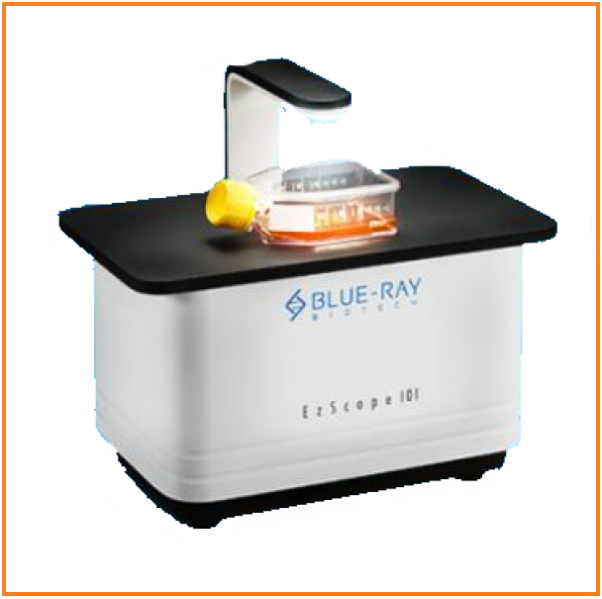
EzScope 101

Live Cell Imaging System – Live Cell Live Show. EzScope 101 is a dedicated live cell imaging system that helps to streamline your research workflow with improved efficiency and productivity, with no more hassles to remove cells from the incubator for observation.

GET BROUCHER

Category:
Description

Description

EzScope 101 is a dedicated live cell imaging system that helps to streamline your research workflow with improved efficiency and productivity, with no more hassles to remove cells from the incubator for observation. EzScope 101 brings 24/7 measurements under precisely controlled conditions in a non-perturbing environment. You can observe the images anytime with walk-away convenience. Up to four samples can be monitored simultaneously in the same incubator.

Features and Benefits

Incubator Live View

Designed to be used inside the incubator, without the need to remove your cells from the incubator to enhance culture quality control.

Minimizes Experimental Variations

Up to four units of EzScope can be set up in the same incubator and controlled by one computer. This enables the monitoring of samples simultaneously and reduces errors caused by environmental variations.

Exceptional Image Quality

Adopts a high contract brightfield optical configuration, coupled with precise motorized focusing, and two interchangeable magnifying objective lenses.

Remote Monitoring of Experiment

Allows flexible remote monitoring of the assay via Windows-based remote desktop software.

Easy Image Editor

Captures and edits images easily with EzCapture software:

● Live preview for up to 4 units of EzScope

● Capture single image or time-lapse series

● Flatfielding correction for even brightfield background

● Time-lapse video output

● Spatial calibration

● Measure and convergence analysis

Specifications

Specifications


Optics Brightfield (transmitted) with white LED
Objective Lens 10x, 20x (optional)
Camera 1.3 MP CMOS Sensor
Image Resolution 1280 x 1024 pixels
Export Formats Tiff (image), AVI (video)
Software EzCapture with snapshot, time-lapse and confluence, etc
Field of View 2.6 x 2.0 mm (10x objective)
Resolution 2 µm/pixel (10x objective), 1 µm/pixel (20x objective)
Live View Frame Rate Up to 8 frames/second
Focusing Motorized
Stage
Manual XY stage (optional) SBS footprint
Labware Holders (optional) 35 mm culture dish and slide, 60 mm culture dish and slide, T-25 culture flask, T-75 culture flask
General
Computer Requirements i3 CPU with 4 GB RAM, Windows 10 OS, i5 CPU with 8 GB RAM, Windows 10 OS for multiple units connection
Connectivity USB 2.0/3.0, up to 4 units
Power Adaptor Input: AC 100-240 V, 50/60 Hz; Output: DC 5 V, 2 A
Dimensions (W x D x H) 225 x 131 x 205 mm
Weight 2.0 kg
Operating Conditions 0°C – 42°C, 5% – 95% RH non-condensing
Certifications CE, RoHS
Reviews (0)

Reviews

There are no reviews yet.

Be the first to review “EzScope 101”

Your email address will not be published. Required fields are marked *

More Products

ezSPR

Leading edge bioanalytical platform for clinical analysis and life sciences.

Benchtop SPR

GET BROUCHER

DILUWEL

Gravimetric Diluter DILUWEL is a rapid, accurate gravimetric diluter for combining solid samples with the correct amount of diluent, automating the laborious task of making and standardizing initial sample dilution.

GET BROUCHER

S700 Rotary Paraffin Microtome

The S700 rotary microtome is a semi-automatic paraffin microtome that embeds tissue in wax and then slices it. Paraffin sections can be used to detect various clinical diseases or help researchers explore tissue and cell levels.

GET BROUCHER

Moxi GO II

Mini Automated Cell Counter Kit Combines the Coulter Principle (for highly-accurate cell counts and exact, volumetric cell sizing) with a 488nm laser and two PMT fluorescence detection channels (for cell health assays, robust CAR-T monitoring, cellular response profiling, and immune profiling).

GET BROCHURE

RED

The Red Tabletop Fluorescence Microscope System by LE.AM Solution delivers powerful, cutting-edge fluorescence imaging in a compact, efficient design suitable for any modern laboratory.

  • Z-Stacking Capabilities: Capture multiple focal planes to generate high-resolution, in-depth 3D visualizations of complex cellular structures.
  • High-Resolution Imaging: Capture intricate cellular details with 12-megapixel resolution and up to 600X magnification.
  • Exceptional Sensitivity: Highly sensitive detectors and a strong optical illumination path easily capture even the faintest fluorescent signals.
  • 4-Channel LED Illumination: Covers a versatile wavelength range from 365nm to 620nm for accurate Biomolecule identification.
  • User-Friendly Operation: Features an intuitive software interface designed for seamless operation and reliable, consistent results.
  • Space-Saving Design: A highly efficient tabletop model that brings state-of-the-art optical innovation right to your lab bench.
  • Built-in Deconvolution: The software includes a specific algorithm to “clear” background noise, providing confocal-like clarity without the confocal price point.
  • Automated Workflow: The motorized Z-stack and one-touch autofocus make it a “smart” microscope compared to traditional manual inverted units

Moxi V

Mini Automated Cell Analyzer The Moxi V provides a combination of volumetric cell sizing (Particle Sizer) with simultaneous fluorescence (Cell Analyzer) to provide the most accurate cell counts, size, and viability in the industry.

GET BROUCHER